Saturday 25 February 2012

Ventricular premature beats (VPBs)

A lady with Romano-Ward syndrome.


Long QT interval

  • The QT interval normally varies with heart rate - becoming shorter at faster rates. It is usually corrected using the cycle length (R-R interval) as shown opposite.
  • normal QTc = 0.42 seconds
Romano-Ward syndrome is an autosomal dominantly inherited form of long QT interval and there is a risk of recurrent ventricular tachycardia, particularly Torsade de Pointes.

Ventricular premature beats (VPBs)

  • 2 ventricular premature beats are also shown in this ECG
  • They are
    • broad
    • occur earlier than normal
    • and are followed by a full compensatory pause (the distance between the normal beats before and after the VPB is equal to twice the normal cycle length).

Ventricular bigeminy


A 50 year old man with chest pain for 24 hours


Ventricular bigeminy

There are also features of an acute inferior myocardial infarction.

Complete Heart Block

A 70 year old man with exercise intolerance.


Complete Heart Block

  • P waves are not conducted to the ventricles because of block at the AV node. The P waves are indicated below and show no relation to the QRS complexes. They 'probe' every part of the ventricular cycle but are never conducted.
  • The ventricles are depolarised by a ventricular escape rhythm.

Ventricular tachycardia

A 45 year old lady with palpitations and history of chronic renal failure

A wide QRS tachycardia is VT until proven otherwise (1). Features suggesting VT include:-

  • evidence of AV dissociation
    • independent P waves (shown by arrows here)
    • capture or fusion beats
    • beat to beat variability of the QRS morphology
  • very wide complexes (> 140 ms)
  • the same morphology in tachycardia as in ventricular ectopics
  • history of ischaemic heart disease
  • absence of any rS, RS or Rs complexes in the chest leads (2)
  • concordance (chest leads all positive or negative)
1) Griffith MJ, Garrat CJ, Mounsey P, Camm AJ. Ventricular tachycardia as the default diagnosis in broad complex tachycardia. Lancet. 1994;343:386-
2) Brugada P, Brugada J, Mont L, et al. A new approach to the differential diagnosis of a regular tachycardia with a wide QRS complex. Circulation. 1991;83:1649-1659

Ventricular tachycardia

A 69 year old man 2 weeks after an inferior myocardial infarction

A wide QRS tachycardia is VT until proven otherwise (1). Features suggesting VT include:-

  • evidence of AV dissociation
    • independent P waves
    • capture or fusion beats
    • beat to beat variability of the QRS morphology (shown here)
  • very wide complexes (> 140 ms)
  • the same morphology in tachycardia as in ventricular ectopics
  • history of ischaemic heart disease
  • absence of any rS, RS or Rs complexes in the chest leads (2)
  • concordance (chest leads all positive or negative)
1) Griffith MJ, Garrat CJ, Mounsey P, Camm AJ. Ventricular tachycardia as the default diagnosis in broad complex tachycardia. Lancet. 1994;343:386-
2) Brugada P, Brugada J, Mont L, et al. A new approach to the differential diagnosis of a regular tachycardia with a wide QRS complex. Circulation. 1991;83:1649-1659

N.B. The computer-aided diagnosis can often be misleading.

Polymorphous ventricular tachycardia (Torsade de pointes).


A 60 year old man with Ischaemic Heart Disease.


  • This is a form of VT where there is usually no difficulty in recognising its ventricular origin.
  • wide QRS complexes with multiple morphologies
  • changing R - R intervals
  • the axis seems to twist about the isoelectric line
  • it is important to recognise this pattern as there are a number of reversible causes
    • heart block
    • hypokalaemia or hypomagnesaemia
    • drugs (e.g. tricyclic antidepressant overdose)
    • congenital long QT syndromes
    • other causes of long QT (e.g. IHD)

Implantable cardioverter defibrillator

A 36 year old lady with recurrent blackouts.
  • Most of this 12-lead recording is polymorphic ventricular tachycardia but, in the rhythm strip, the large deflection (arrowed) is the defibrillator discharging.
  • Following the defibrillation a dual chamber pacemaker can be seen.
OK so I cheated a little with this one as the odds of catching this on a 12-lead ECG recording are very slim indeed. This is a reconstructed 12-lead recording from an electrophysiology study testing the device after placement.

Ventricular fibrillation

A 60 year old man with 2 hours of "crushing" chest pain suddenly collapses.
  • bizarre, irregular, random waveform
  • no clearly identifiable QRS complexes or P waves
  • wandering baseline
  • A 12 lead of Ventricular fibrillation should not usually be taken ... for obvious reasons. Instead of continuing to record the ECG you should check the patient's pulse and reach for the defibrillator!

Ventricular pacemaker

A 72 year old man with a permanent pacemaker.

  • pacing spikes (best seen here in V4 - V6) will be seen - they may be subtle
  • the paced QRS complexes are abnormally wide
In this example the pacemaker starts when there is a long R - R interval following a blocked atrial premature beat (arrowed in figure below). Sinus rhythm takes over again later in the rhythm strip.

Implantable cardioverter defibrillator

A 36 year old lady with recurrent blackouts.


  • Most of this 12-lead recording is polymorphic ventricular tachycardia but, in the rhythm strip, the large deflection (arrowed) is the defibrillator discharging.
  • Following the defibrillation a dual chamber pacemaker can be seen.
OK so I cheated a little with this one as the odds of catching this on a 12-lead ECG recording are very slim indeed. This is a reconstructed 12-lead recording from an electrophysiology study testing the device after placement.

Wolf-Parkinson-White syndrome

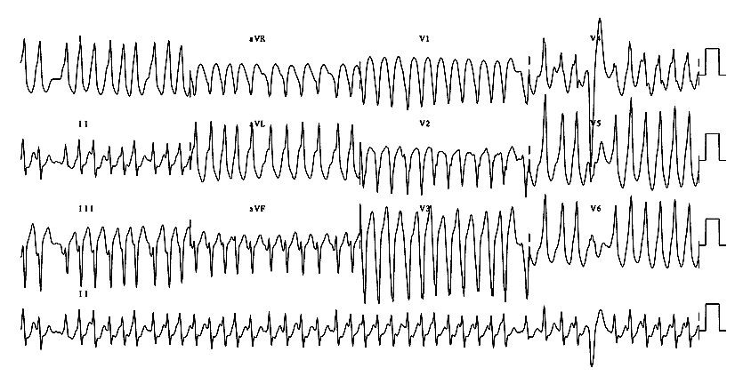
A 25 year old man with bouts of tachycardia.

  • short PR interval, less than 3 small squares (120 ms)
  • slurred upstroke to the QRS indicating pre-excitation (delta wave)
  • broad QRS
  • secondary ST and T wave changes

Localising the accessory pathway

An accessory pathway, bundle of Kent, exists between atria and ventricles and causes early depolarisation of the ventricle. The location of the pathway may be deduced as follows:-
  LOCATION                   V1   V2   QRS axis
left posteroseptal (type A)  +ve  +ve  left
right lateral      (type B)  -ve  -ve  left
left lateral       (type C)  +ve  +ve  inferior (90 degrees)
right posteroseptal          -ve  -ve  left
anteroseptal                 -ve  -ve  normal

Wolf-Parkinson-White syndrome

A 23 year old man with epsiodes of tachycardia.
  • short PR interval, less than 3 small squares (120 ms)
  • slurred upstroke to the QRS indicating pre-excitation (delta wave)
  • broad QRS
  • secondary ST and T wave changes

Localising the accessory pathway

An accessory pathway, bundle of Kent, exists between atria and ventricles and causes early depolarisation of the ventricle. The location of the pathway may be deduced as follows:-
  LOCATION                   V1   V2   QRS axis
left posteroseptal (type A)  +ve  +ve  left
right lateral      (type B)  -ve  -ve  left
left lateral       (type C)  +ve  +ve  inferior (90 degrees)
right posteroseptal          -ve  -ve  left
anteroseptal                 -ve  -ve  normal

Wolf-Parkinson-White syndrome with atrial fibrillation

A 47 year old man with a long history of palpitations and, lately, blackouts.

  • irregularly irregular, wide complex tachycardia
  • impulses from the atria are conducted to the ventricles via either
    • both the AV node and accessory pathway producing a broad fusion complex
    • or just the AV node producing a narrow complex (without a delta wave)
    • or just the accessory pathway producing a very broad 'pure' delta wave
  • people who develop this rhythm and have very short R - R intervals are at higher risk of VF
  • Wolf-Parkinson-White syndrome with atrial fibrillation

    A 23 year old man with episodes of palpitations.
    • irregularly irregular, wide complex tachycardia
    • impulses from the atria are conducted to the ventricles via either
      • both the AV node and accessory pathway producing a broad fusion complex
      • or just the AV node producing a narrow complex (without a delta wave)
      • or just the accessory pathway producing a very broad 'pure' delta wave
    • people who develop this rhythm and have very short R - R intervals are at higher risk of VF